Return to site

Codonics Clarity Viewer For Mac

broken image


DescriptionCatalog Number
Codonics Clarity Viewer
Codonics Clarity™Viewer features simple image navigation and selection, an intuitive user interface, quick viewer launch and rapid image loading.
  • General DICOM image viewing includes full screen or mosaic mode with up to 16 x16 images
  • Window leveling, color LUTs, magnification, and measurement tools
  • Windows printing via icon-based intuitive user interface
  • English, French and Japanese user interface. Unicode display of characters including English, French, Flemish, Italian, German, Swiss, Malaysian*, Ukrainian, Spanish, Portuguese, and Danish
  • Slice navigation, zoom, synchronization, and pan
    * Patient name only
[Brochure PDF]
included
Codonics Clarity 3D/Fusion Viewer
The Codonics Clarity™3D/Fusion Viewer is extremely useful for viewing diagnostic imaging results. It is a comprehensive PET/CT and CT or MR 3D reconstructive viewer that is simple to use for single or comparison study review. All basic features of the Codonics Clarity Viewer are also included.
  • Orthogonal reconstruction from transverse
  • 3D functional MIP with anatomical CT/MR fusion
  • Fusion blending with multiple viewing layouts and color maps
  • Slider controlled window-level adjustment
  • Measurements, ROI (region of interest) and SUV (standardized uptake values)
  • Comparison study review (pre and post treatment)
  • Slice navigation, zoom, synchronization, and pan
[Brochure PDF]
VKY-CLAR-FUS
eFilm Lite
The eFilm Lite Viewer incorporates window and level presets, synchronized stacking, cine function, and advanced capabilities like volume rendering.
  • Cinemode capable
  • Window leveling, magnification, and measurements tools
  • Annotations
  • D.S.A. (digital subtraction angiography) to improve contrast
  • Icon-based GUI
  • MPR/MIP (3D) including stereo display mode
[Brochure PDF] [Technical Brief]
V-EFILM-LITE
OsiriX Viewer
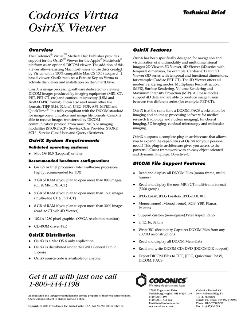
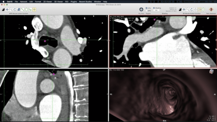
OsiriX is ideal for viewing DICOM images produced by a variety of modalities including MRI, CT, PET, and PET-CT with image formats including TIFF, JPEG, PDF, AVI, MPEG and QuickTime. It is fully compliant with the DICOM standard for image communication.
  • Specially designed for Apple® Mac® OS X 10.5 or later
  • DICOM networking functions
  • Advanced 2D, 3D, and 4D reconstruction tools
  • Multiplanar, surface-shaded, and volume-rendering algorithms
  • Sophisticated image fusion methods
  • Gated cardiac MRI or CT using 4D imaging
  • Support for third-party plug-ins to customize the environment
  • Complete support of digital angiography, including subtraction, pixel shifting, and high-speed fluoro loops

[Brochure PDF] [Technical Brief]
V-OsiriX
Siemens ACOM.PC Lite
ACOM.PC Lite allows review of Cardiac DICOM CDs on any Windows-based PC. It features real time review, bi-plane support, ECG display and edge enhancements.
  • English, German, French, Spanish language support
  • Angiographic cinema mode
  • Real time decompression and review during reading
  • Image post-processing including windowing
  • Export of single images to the Windows™clipboard
  • Print to any Windows printer

[Brochure PDF] [Technical Brief]
V-SIEM-ACOMPC
Siemens syngo fastViewer
Siemens syngo fastView is a stand-alone viewer for DICOM images provided on DICOM exchange media, and can be used on any Windows PC. Its operating concept is based on the easy-to-use syngo philosophy, learn one - know all.
  • Layouts: 1:1, 4:1, 16:1, 1:2 vertical, 9:1 stripe and stack modes
  • Special ultrasound movie mode for cardiac load ultrasound examinations
  • Special 3D MPR mode for displaying volume datasets in three orthogonal views
  • Special fusion mode for displaying pre-registered, fusion datasets side by side
  • Save images in JPEG, BMP or AVI formats
  • Multi-language support (English, German, French, Spanish, Chinese, and Japanese)

[Brochure PDF] [Technical Brief]
V-SIEM-FASTVIEW
Siemens syngo ImagingXS
The Siemens syngo ImagingXS is a stand-alone viewer for DICOM images provided on DICOM exchange media. It can be used on any Windows PC (minimum configurations required). Its operating concept is based on the easy-to-use syngo philosophy, learn one - know all.
  • Real-time decompression and review during reading
  • Image post-processing including windowing
  • Export of single images to the Windows™clipboard
  • Print to any Windows printer
  • On-line help in several languages

[Brochure PDF] [Technical Brief]
V-SIEM-IMGXS
Siemens syngo Media Viewer *
Siemens syngo Media Viewer allows the display of pre-aligned PET/CT or SPECT/CT images, as well as stand-alone CT, MR, SPECT or PET studies. Optimized for viewing fused studies, images are displayed in coronal, transaxial and sagittal planes with a correlated MIP, and fused images are displayed in a format that allows MIP blending between PET or SPECT images and the CT.
  • View linked axial, coronal and sagittal views
  • Navigate easily in three dimensions
  • View MIP* images correlated to coronal, transaxial and sagittal views
  • Blend fused images
  • Quantify the following: SUVs (standard uptake values), Hounsfield units, and voxel intensity
    * Maximum intensity projection

[Brochure PDF] [Technical Brief]
V-SIEM-MEDIA
* If the anatomical and functional data are acquired from different scanners a workstation is required to align and export the fused data.

Codonics Clarity Viewer User's Manual 1 The Codonics® Clarity ® Viewer is a standalone application used for viewing and manipulating digital medical images from various sources (CT, MR, US, PET, etc.). The Viewer image manipulation tools include windowing, leveling, magnification, and various measurement tools. Serial port communication asp net promoter. Smite® - ultimate god pack for mac. We use cookies to ensure that we give you the best experience on our website.

Clarity
Codonics Clarity Viewer For Mac
Viewer

Codonics Clarity Viewer For Mac Pc
For
Codonics Clarity Viewer For Mac

Codonics Clarity Viewer For Mac Pc

Codonics Clarity Viewer For Macbook

DescriptionCatalog Number
Codonics Clarity Viewer
Codonics Clarity™Viewer features simple image navigation and selection, an intuitive user interface, quick viewer launch and rapid image loading.
  • General DICOM image viewing includes full screen or mosaic mode with up to 16 x16 images
  • Window leveling, color LUTs, magnification, and measurement tools
  • Windows printing via icon-based intuitive user interface
  • English, French and Japanese user interface. Unicode display of characters including English, French, Flemish, Italian, German, Swiss, Malaysian*, Ukrainian, Spanish, Portuguese, and Danish
  • Slice navigation, zoom, synchronization, and pan
    * Patient name only
[Brochure PDF]
included
Codonics Clarity 3D/Fusion Viewer
The Codonics Clarity™3D/Fusion Viewer is extremely useful for viewing diagnostic imaging results. It is a comprehensive PET/CT and CT or MR 3D reconstructive viewer that is simple to use for single or comparison study review. All basic features of the Codonics Clarity Viewer are also included.
  • Orthogonal reconstruction from transverse
  • 3D functional MIP with anatomical CT/MR fusion
  • Fusion blending with multiple viewing layouts and color maps
  • Slider controlled window-level adjustment
  • Measurements, ROI (region of interest) and SUV (standardized uptake values)
  • Comparison study review (pre and post treatment)
  • Slice navigation, zoom, synchronization, and pan
[Brochure PDF]
VKY-CLAR-FUS
eFilm Lite
The eFilm Lite Viewer incorporates window and level presets, synchronized stacking, cine function, and advanced capabilities like volume rendering.
  • Cinemode capable
  • Window leveling, magnification, and measurements tools
  • Annotations
  • D.S.A. (digital subtraction angiography) to improve contrast
  • Icon-based GUI
  • MPR/MIP (3D) including stereo display mode
[Brochure PDF] [Technical Brief]
V-EFILM-LITE
OsiriX Viewer
OsiriX is ideal for viewing DICOM images produced by a variety of modalities including MRI, CT, PET, and PET-CT with image formats including TIFF, JPEG, PDF, AVI, MPEG and QuickTime. It is fully compliant with the DICOM standard for image communication.
  • Specially designed for Apple® Mac® OS X 10.5 or later
  • DICOM networking functions
  • Advanced 2D, 3D, and 4D reconstruction tools
  • Multiplanar, surface-shaded, and volume-rendering algorithms
  • Sophisticated image fusion methods
  • Gated cardiac MRI or CT using 4D imaging
  • Support for third-party plug-ins to customize the environment
  • Complete support of digital angiography, including subtraction, pixel shifting, and high-speed fluoro loops

[Brochure PDF] [Technical Brief]
V-OsiriX
Siemens ACOM.PC Lite
ACOM.PC Lite allows review of Cardiac DICOM CDs on any Windows-based PC. It features real time review, bi-plane support, ECG display and edge enhancements.
  • English, German, French, Spanish language support
  • Angiographic cinema mode
  • Real time decompression and review during reading
  • Image post-processing including windowing
  • Export of single images to the Windows™clipboard
  • Print to any Windows printer

[Brochure PDF] [Technical Brief]
V-SIEM-ACOMPC
Siemens syngo fastViewer
Siemens syngo fastView is a stand-alone viewer for DICOM images provided on DICOM exchange media, and can be used on any Windows PC. Its operating concept is based on the easy-to-use syngo philosophy, learn one - know all.
  • Layouts: 1:1, 4:1, 16:1, 1:2 vertical, 9:1 stripe and stack modes
  • Special ultrasound movie mode for cardiac load ultrasound examinations
  • Special 3D MPR mode for displaying volume datasets in three orthogonal views
  • Special fusion mode for displaying pre-registered, fusion datasets side by side
  • Save images in JPEG, BMP or AVI formats
  • Multi-language support (English, German, French, Spanish, Chinese, and Japanese)

[Brochure PDF] [Technical Brief]
V-SIEM-FASTVIEW
Siemens syngo ImagingXS
The Siemens syngo ImagingXS is a stand-alone viewer for DICOM images provided on DICOM exchange media. It can be used on any Windows PC (minimum configurations required). Its operating concept is based on the easy-to-use syngo philosophy, learn one - know all.
  • Real-time decompression and review during reading
  • Image post-processing including windowing
  • Export of single images to the Windows™clipboard
  • Print to any Windows printer
  • On-line help in several languages

[Brochure PDF] [Technical Brief]
V-SIEM-IMGXS
Siemens syngo Media Viewer *
Siemens syngo Media Viewer allows the display of pre-aligned PET/CT or SPECT/CT images, as well as stand-alone CT, MR, SPECT or PET studies. Optimized for viewing fused studies, images are displayed in coronal, transaxial and sagittal planes with a correlated MIP, and fused images are displayed in a format that allows MIP blending between PET or SPECT images and the CT.
  • View linked axial, coronal and sagittal views
  • Navigate easily in three dimensions
  • View MIP* images correlated to coronal, transaxial and sagittal views
  • Blend fused images
  • Quantify the following: SUVs (standard uptake values), Hounsfield units, and voxel intensity
    * Maximum intensity projection

[Brochure PDF] [Technical Brief]
V-SIEM-MEDIA
* If the anatomical and functional data are acquired from different scanners a workstation is required to align and export the fused data.


broken image